- سبدخرید خالی است.
- ادامه خرید

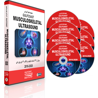
Gulfcoast Musculoskeletal Ultrasound
شاید شما این را نیز دوست داشته باشید…
توضیحات محصول
DVD 1:
Advanced Ultrasound Techniques for the Foot and Ankle:
- LATERAL:
- Calcaneofibular Ligament
- Static and Dynamic
- Subtalar Joint
- Inframalleolar Peroneal
- Distal Peroneals
- Osperoneum
- POSTEROLATERAL:
- Peroneals
- Superior Peroneal Retinaculum
- Low Lying Brevis or Quartus Muscle Belly
- ANTEROLATERAL:
- Anterior Talofibular Ligament
- Static and Dynamic
- AITFL, Static and Dynamic
- Anterolateral Gutter
- POSTERIOR:
- Achilles
- Kager’s Fat Pad
- Retrocalcaneal Bursa Impingement
- Os Trigom Injections
- Subtalar Joint
- MEDIAL:
- Medial Tendon’s
- Identify common Arthritic Joints and Setup Injections for the Midfoot
- Plantar and Dorsal Evaluations of the Forefoot / Morton’s Neuroma
Advanced Ultrasound-Guided Procedures for Rehabilitation Medicine:
- PRP to Atrophied Lumbar Multifidi
- Spine Pain Treatment Options
- Longissimus Muscle Strain
- Rectus Abdominus Strain
- Hamstring Strain
- Ultrasound-Guided ACL Injection
- LCL / RCL Defects
- Regenerative Medicine Applications
- Pitfalls and Benefits of Ultrasound-guided MSK Intervention Techniques
Bone Marrow _ Lipoaspirate – General Principles _ Practical Applications:
- Biologic Therapies / Treatments
- Mesenchymal Stem Cells
- How is BMC Different from PRP
- IRAP or Orthokine
- Lipo-Aspiration
- Safe Zones of the Ilium
- Stromal Vascular Fraction
Cases in Trauma Ultrasound
- Bladder Rupture
- Splenic Injury
- Pneumothorax
- Hemothorax
- Stab Wound to the Chest
- Pericardial Effusion
- Foreign Body in Hand & Back
- Gun Shot Wound to Leg
- Traumatic Tendon Injury
- Fracture
- Shoulder Dislocation
- Traumatic Vision Loss
- Head Injury with Vision Loss
Dynamic Musculoskeletal Ultrasound Imaging:
- HOULDER:
- Biceps Brachii Tendon Subluxation/Dislocation
- Impingement Syndrome
- Adhesive Capsulitis
- ELBOW:
- Ulnar Nerve Dislocation
- Snapping Triceps Syndrome
- Ulnar Collateral Ligament Partial Tear, Ulnar Collateral Ligament Laxity
- Ulnar Collateral Ligament Complete Tear
- WRIST & HAND:
- Extensor Carpi Ulnaris Dislocation
- Boxer Knuckle
- Pulley Tear
- Trigger Finger
- Ganglion Cyst
- Gamekeeper’s Thumb
- HIP & THIGH:
- Snapping Hip Syndrome (Iliopsoas; Iliotibial Tract)
- KNEE:
- Patellar Tendon Full Thickness Tear
- Meniscal Displacement
- Bucket Handle Tear Medial Meniscus
- Intra-Articular Body
- Patellar Clunk Syndrome
- Snapping Semitendinosus Tendon
- Snapping Sartorius
- ANKLE/FOOT::
- Peroneal Tendon Dislocation/Subluxation
- Tendon Impingement
- Muscle Hernia
- Achilles Tendon
- Morton Neuroma
- SOFT TISSUES:
- Slipping Rib Syndrome
Soft-Tissue and MSK Sonography in the Pediatric Patient
- Cellulitis
- Necrotizing Fasciitis
- Abscess
- Foreign Body Localization/Removal
- Fracture Evaluation and Reduction
- Tendon Injuries
- Nerve Blocks
- Joint Effusion: Hip & Knee
DVD 2:
Focused Assessment with Sonography in Trauma:
- Ultrasound Evaluation of the Hemoperitoneum
- Ultrasound Evaluation of the Hemopericardium
- Ultrasound Evaluation of the Hemothorax
- Ultrasound Evaluation of the Pneumothorax
Lower Extremity Musculoskeletal Ultrasound Protocols
- Normal Ultrasound Anatomy and Protocols for the Knee, and Ankle/Foot
- Normal Ultrasound Characteristics for the Knee, and Ankle/Foot
- Ankle and Foot Exam:
- Extensor Tendons
- Extensor Digitorum Longus Tendon
- Tibio-Talar Joint
- Medial Malleolus
- Posterior Tibial Tendon
- Posterior Tibial Nerve
- Flexor Hallucis Longus
- Lateral Malleolus
- Anterior Talo-Fibular Ligament
- Tibio-Fibular Ligament
- Achilles Tendon
- Plantar Fascia
- Knee Exam:
- Quadriceps Tendon
- Patellar Tendon
- Medial Collateral Ligament
- Lateral Collateral Ligament
- Popliteal Fossa
- Posterior Medial Meniscus
- Posterior Lateral Meniscus
Musculoskeletal Ultrasound Evaluation of the Hip:
- Joint abnormalities: hip effusion, labral tear, paralabral cyst, femoro-acetabular impingement, hip arthroplasty
- Bursal Pathology: trochanteric pain syndrome, trochanteric bursitis, iliopsoas bursal fluid
- Muscle and tendon injury: acute muscle and tendon injury, tendinosis, semimembranosus tear, sports hernia, complete tear adductor longus, rectus femoris tear, calcific tendinosis, iliopsoas hemorrhage
- Snapping Hip syndrome
- Miscellaneous pathology: Morel-Lavallée lesion; Acute and Chronic cellulitis
- Soft-tissue abscess
- Inflammatory myositis
- Lymph node evaluation
- Soft-tissue myxoma
- Soft-tissue sarcoma
Musculoskeletal Ultrasound for Postoperative Applications
- Rotator Cuff Repair Status / Re-Rupture
- Causes of Impingement / Pain
- Persistent Interstitial Tears
- Tendinosis
- Bursitis
- Post-Operative Scar Tissue
- POST-OPERATIVE HIP:
- Total Hip Arthroplasty / Labral Repair
- Pseudo-Tumor Evaluation
- Aspiration
- Infection
- Gluteal Tendon Repair
- POST OPERATIVE KNEE:
- Partial or Total Knee Arthroplasty
- Quadricep Tendinosis
- Scar Tissue
- Patellar Tendinosis
- Infrapatellar Band
- Saphenous Nerve Block
- Peroneal Nerve Palsy
- Evaluation for Effusion / Aspiration
- POST-OPERATIVE FOOT/ANKLE:
- Peroneal Pain
- Sural Nerve Evaluation
- Hydrodissection
- Adhesive Capsulitis
- Ligamentous/Capsular Arthrofibrosis Debridement
Ultrasound-Guided Peripheral Nerve Injections in an MSK Practice
- Indications for Peripheral Nerve Injections
- Transducer Selection and Image Optimization
- Nerve Anatomy
- Ultrasound Characteristics
- Pre-Procedural Considerations
- Needle Selection and Injection Techniques
- Upper Extremity Ultrasound-Guided Procedures: Suprascapular Nerve Injection, Neuropathy of the Upper Extremity – Median Nerve, Radial Nerve, Radial Tunnel Syndrome, Posterior Interosseous Nerve Entrapment, Radial Nerve Branches, Cubital Tunnel Syndrome, Cubital Tunnel Injection, Ulnar Nerve Block, Carpal Tunnel Injection, Ulnar Neuropathy at Guyon’s Canal, Peripheral Nerve Tumor, and Median Nerve Hamartoma.
- Lower Extremity Peripheral Nerve Injections: Stump Neuromas, Lateral Femoral Cutaneous Nerve Block, Common Peroneal Neuropathy, Common Peroneal Nerve Hydro-Dissection, Tibia-Fibular Joint Ganglia, Superficial Peroneal Neuropathy, Deep Peroneal Neuropathy, Tarsal Tunnel Syndrome, tibial Nerve, Baxter’s Neuropathy, Lateral Plantar Nerve Injection, and Morton’s Neuroma Injection
DVD 3
Ultrasound Evaluation of Knee Pathology:
- Pathology of the Anterior, Medial, Lateral and Posterior Knee
- ANTERIOR: Quadriceps & Patellar Tendons, Patella, and Fat Pads
- Joint Effusion, Synovitis, Loose Bodies, Plica
- Bursae: Pre-Patellar, Deep and Superficial Infrapatellar
- MEDIAL: MCL, Pes Anserine, Medial Meniscus, MPFL
- LATERAL: IT Band, Lateral Collateral Ligament, Popliteus, Biceps Femoris, Lateral Meniscus
- POSTERIOR: Peroneal Nerve, Baker’s Cyst, Neurovascular Bundle, PCL
Ultrasound Evaluation of Shoulder Pathology
- Rotator cuff tears, abnormalities
- Pathogenesis
- Supraspinatus and insertion partial thickness tears
- Articular partial-thickness tears
- Bursal partial-thickness tears
- Large full-thickness tears,
- Massive full-thickness tears
- Intrasubstance tears
- Tendinosis
- Fatty infiltration and muscle atrophy
- Tendon volume loss
- Joint and bursal effusions
- Secondary findings of rotator cuff tears
- Pitfalls in rotator cuff sonography
- Other miscellaneous cuff pathology
Ultrasound Evaluation of the Foot and Ankle
- Anterior Foot and Ankle Normal Anatomy
- Medial and Lateral Ankle Normal Anatomy
- Posterior Ankle and Plantar Foot Normal Anatomy
- Patient Positioning and Scan Techniques
- Commonly Seen Pathology
- Effusion
- Tendinopathy
- Spring Ligament
- Tarsal Tunnel Syndrome
- Flexor Hallucis Longus Tendinitis
- Peroneal Tendinopathy
- Peroneal Tendon Subluxation/Dislocation
- Peroneus Quartus
- ATFL Tear
- Achilles Tendinopathy
- Achilles Tendinopathy and Rupture
- Retrocalcaneal Bursitis
- Plantar Fasciitis
- Midfoot Degenerative Joint Disease
- Morton’s Neuroma
- Hallux Rigidus
Ultrasound Evaluation of the Hand and Wrist
- Hand and Wrist Anatomy
- Probe Control
- Normal Sonographic Characteristics of the Hand and Wrist
- Minor Pathologies
- Dorsal Wrist
- Ulnar Wrist
- Radial Wrist
- Volar Wrist
- MCP Joint
- PIP Joint
- Scan Demonstration
DVD 4
Ultrasound Evaluation of Elbow Pathology
- Joint Effusion and Bursa
- Biceps and Triceps Tendon Abnormalities
- Epicondylitis
- Ulnar Collateral Ligament
- Cubital Tunnel
Ultrasound Evaluation of Intima-Media Thickness
- What is Intima Media Thickness?
- Why is IMT Important?
- How to Perform IMT Measurements
- How to Apply IMT Measurements in the Clinical Setting
Ultrasound Evaluation of the Normal Elbow
- Anterior Elbow and Joints
- Pronator Window
- Distal Biceps
- Biceps Tendon
- Medial Elbow
- Common Flexor
- Ulnar Nerve
- Triceps
- Radial Collateral Ligament
- Radial Nerve
- Annular Ligament
- Elbow Scan Demonstration
Ultrasound Evaluation of the Normal Knee
- Normal Knee Anatomy
- Ultrasound Characteristics of Normal Knee Anatomy
- Scan Techniques to Evaluate the Anterior, Medial, Lateral, and Posterior Knee
- Scan Techniques and Protocol for Evaluating the Knee
- Limitations and Pitfalls Associated with the Ultrasound Evaluation of the Knee
- Scan Demonstration
Ultrasound Evaluation of the Pediatric Hip
- Indications for Performing Hip Ultrasounds
- Embryology
- Normal Hip Anatomy
- Ultrasound Scanning Techniques and Protocols
- Two-Step Dynamic Technique
- Diagnostic Criteria for Developmental Dysplasia of the Hip (DDH)
- Lax Hip, Subluxable Hip, Dislocated Hip
- Scan Protocols for Scanning Infants in Pavlik Harness
- Treatment Sonography Protocol
Ultrasound Evaluation of the Shoulder
- Rotator cuff tears, abnormalities
- Pathogenesis
- Supraspinatus and insertion partial thickness tears
- Articular partial-thickness tears
- Bursal partial-thickness tears
- Large full-thickness tears,
- Massive full-thickness tears
- Intrasubstance tears
- Tendinosis
- Fatty infiltration and muscle atrophy
- Tendon volume loss
- Joint and bursal effusions
- Secondary findings of rotator cuff tears
- Pitfalls in rotator cuff sonography
- Other miscellaneous cuff pathology
DVD 5
Advanced Peripheral Nerve Applications – Diagnosis and Treatment Options
- Evaluation of Focal Neuropathies
- Neurapraxia, Axonotmesis, and Neurotmesis
- Nerve Trauma
- Principles of Imaging Peripheral Nerves
- Nerve Echotexture
- Imaging Strategies
- Measurement of Cross-Sectional Area
- Post-surgical Evaluation
Musculoskeletal Imaging Fundamentals and Tissue Characterization
Musculoskeletal Ultrasound Evaluation of Tumors and Masses
- Tumor and Pseudotumor
- Tendon Tear with Retraction
- Muscle Hernia
- Anomalous Muscle
- Rheumatoid Nodule
- Anatomic Location: Joint Recess, Bursa, Tendon, Lymph Node, Ganglion Subcutaneous or Other
- Pigmented Villonodular Synovitis
- Baker’s Cyst
- Bicipitoradial Bursitis
- Gout
- Reactive Lymph Node
- B Cell Lymphoma
- Angiosarcoma Metastasis
- Ganglion Cysts
- Parameniscal Cysts
- Subcutaneous Masses: Fat Necrosis, Epidermal Inclusion Cyst, Lipoma, Fat Necrosis
- Other Malignant Masses: Sarcoma, Metastasis, Melanoma
Musculoskeletal Ultrasound Peripheral Nerve Entrapment
- Peripheral Nerve Entrapment
- Carpal Tunnel Syndrome
- Median and Ulnar Nerve Entrapment Syndromes
- Radial and Suprascapular Nerve Entrapments; Labral Cyst
- Peroneal, Tibial and Interdigital Nerve Entrapment Syndromes
Reducing Your Risk for Occupational Injury
Reducing Your Risk for Occupational Injury Training Video describes the characteristics of musculoskeletal strain injury (MSI) among sonographers, and its impact on the worker and employer. Participants will be presented with the information to assist in identifying risk factors in their work environment. Additionally, the value of being proactive in the prevention of occupational injury will be discussed
Trauma Ultrasound for the Sonographer
- FAST Exam
- Lung Ultrasound: Pneumothorax, Pleural Effusion, and Pulmonary Edema
- Evaluation of the Source of Shock and Hypotension
- Use of Ultrasound in Cardiac Arrest
- Ocular Trauma
- Use of Ultrasound in Fracture Assessment
DVD 6
Ultrasound Evaluation of the Spine _ Injection Techniques
- Lumbar Epidural Steroid Injections
- Caudal Injections
- SI joint Injections
- Cervical Spine Interventions
- Benefits of Prolotherapy
- Treatment Considerations for the Lumbar and Cervical Spine
Ultrasound-Guided Injections in Musculoskeletal Medicine
- Injection Techniques
- Joint Imaging
- Tendon Sheath Imaging
- Bursa Imaging
- Cyst Imaging
- Summary
Ultrasound-Guided Regional Anesthesia – Upper Extremities
- Imaging Fundamentals
- Indications and Applications for Ultrasound-Guided Regional Anesthesia
- Upper Extremity Nerve Blocks: Anatomy, US Scanning and Injection Techniques
- Interscalene
- Supraclavicular
- Infraclavicular
- Axillary
- Musculocutaneous
- Forearm: Radial, Ulnar, Median
- Truncal Blocks
- PECS 1
- PECCS 11
- Subcostal TAP
- Classical TAP
- Quadratus Lumborum Block
- Lower Extremity Nerve Blocks: Anatomy US Scanning and Injection Techniques
- Femoral
- Fascia Iliaca Block
- Saphenous/Adductor Canal Block
- Sciatic (Popliteal & Subgluteal)
- Ankle Blocks
- Scan Demonstrations
Use of Ultrasound in Rheumatology Applications
- Prevalence of Disease in the U.S.
- Rheumatoid Arthritis: Erosions, Synovitis, Tenosynovitis, Treatment Response
- Spondyloarthritis: Synovitis, Tenosynovitis, Peritendinitis, Enthesitis, Erosions, New Bone Formation
- Crystal Arthritis: Sonographic Features of Gout
- Osteoarthritis: Synovitis, Systemic Inflammatory Mediators, Low-Grade Inflammation, Articular Cartilage, Basic Calcium Phosphate Crystals
DVD 7
Gulfcoast: MSK Ultrasound Guided PRP: General Principles and Applications
- PRP: What is it?
- PRP: What’s in it?
- PRP: Growth Factors.
- PRP: How does it work and Why use it?
- PRP: What does it treat?
- Advantages and Criticisms.
- PRP: The Harvest and Preparation.
- What about Anesthetic?
- PRP Evidence
- PRP Applications: Patellar Tendon, Chronic Jumpers Knee, Lateral & Medial Epicondyle; Posterior SI Ligaments; Gluteus Medius; Proximal Hamstring; Mean Nerve Hydrodisection; Supraspinatus; MCL
Ultrasound-Guided Prolotherapy – General Principles
- History of Prolotherapy
- Mechanisms of Prolotherapy
- Injection Solutions
- Treatment Principles and Techniques
- Outcomes of Prolotherapy
- Injection Techniques
- Diagnostic Considerations for Musculoskeletal Abnormalities
- The Role of Prolotherapy
- Conditions that respond well to the use of Prolotherapy
Ultrasound-Guided Regional Anesthesia – Lower Extremities
Ultrasound-Guided Upper Extremity Nerve Blocks for Emergency Medicine
- Overview of Needle Guidance Basics
- Indications and Applications for Ultrasound-Guided Nerve Blocks
- Ultrasound-Guided Nerve Blocks for the Upper Extremity: Anatomic Territories, Interscalene Block, Supraclavicular Brachial Plexus Block, Axillary Plexus Block, Forearm Nerve Blocks, and Cervical Plexus Block
- Upper Extremity Nerve Block Case Studies
- Billing and Coding
Upper Extremity Musculoskeletal Ultrasound Protocols
-
- Normal ultrasound anatomy and protocols for the shoulder, elbow, hand/wrist
- Normal ultrasound characteristics for the shoulder, elbow, hand/wrist
- Shoulder Exam:
-
- Biceps Tendon
- Subscapularis Tendon
- Rotator Cuff
- Supraspinatus Tendon
- Rotator Cuff Interval
- Infraspinatus Tendon
- Acromioclavicular Joint
- Glenohumeral joint
- Shoulder Impingement Syndrome
-
- Elbow Exams:
- Anterior Elbow Views (Short and Long Axis)
- Medial Epicondyle
- Posterior Elbow Views (Short and Long Axis)
- Ulnar Nerve
- Ulnar Nerve Subluxation
- Wrist and Hand Exam:
- Median Nerve


