

2025 Fetal Echocardiography: Normal and Abnormal Hearts | Fetal Heart Ultrasound Course – Simabaan
2025 Fetal Echocardiography: Normal and Abnormal Hearts
The Most Comprehensive Diagnostic Course in Fetal Cardiac Ultrasound
If you regularly deal with high-risk pregnancies, suspected fetal heart disease, or complex fetal scans,
fetal echocardiography is not just an extra skill for you – it’s a critical tool for life-changing decisions during pregnancy.
2025 Fetal Echocardiography: Normal and Abnormal Hearts is one of the most up-to-date and reputable educational programs in this field. It is built around a complete evaluation of the normal fetal heart and the diagnosis of congenital heart disease (CHD).
Taught by leading experts in Maternal–Fetal Medicine and Pediatric Cardiology, this course provides a systematic, practical, case-based framework for:
-
Structural assessment of the fetal heart
-
Recognition of abnormalities
-
Interpretation of images and hemodynamic findings
Why Fetal Echocardiography Matters Even More in 2025
With advances in imaging technology, expectations from obstetricians, MFM specialists and pediatric cardiologists go far beyond “just seeing a four-chamber view.”
Today, you are expected to:
-
Define a truly normal fetal heart with precision
-
Detect abnormalities early and accurately
-
Design a logical management and follow-up plan for the pregnancy and the newborn
This course is designed exactly for that need:
a practical, high-yield guide for anyone who wants to go beyond a “basic fetal heart scan” and move closer to the level of a fetal cardiac specialist.
📌 1. Essentials of Fetal Cardiac Anatomy
Precise fetal cardiac anatomy – the foundation of every accurate diagnosis
The first module focuses on complete fetal cardiac anatomy in the second and third trimesters. You will become familiar with:
-
The main structural components of the fetal heart
-
Chambers, valves, septa and great vessels
-
How these structures relate to each other in standard ultrasound views
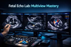
Standard imaging planes are reviewed step by step, including:
-
Four-chamber view (4CV)
-
Outflow tract views (LVOT, RVOT)
-
Three-vessel view and three-vessel trachea view (3VV / 3VTV)
The goal is to move from a “global” look at the heart to a structured, reproducible anatomical assessment that you can apply in every scan.
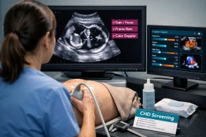
2. Fetal Echocardiography Techniques & Image Optimization
Image quality is half of the diagnosis
This module focuses on practical scanning techniques and machine optimization for fetal cardiac imaging:
-
Best probe positions and angles to obtain ideal views
-
Key machine settings to improve resolution, depth, gain and focus
-
How to deal with common challenges:
-
Fetal movement
-
Suboptimal fetal position
-
High maternal BMI
-
Technical limitations
-
-
Frequent scanning errors – and how to avoid them
By the end of this section, you’ll know how to get the best possible image quality from the machine you already use, which directly translates into better diagnostic confidence.
⚠️ 3. Congenital Heart Diseases (CHD): Identification & Diagnosis
From a normal heart to the most complex CHDs
One of the most important parts of the course is a detailed review of common and clinically significant congenital heart diseases. This includes:
-
Septal defects (ASD, VSD) and their imaging patterns
-
Outflow tract abnormalities, asymmetry, malalignment and related anomalies
-
Complex lesions such as:
-
HLHS – Hypoplastic Left Heart Syndrome
-
TGA – Transposition of the Great Arteries
-
TOF – Tetralogy of Fallot
-
and other major CHDs
-
-
Assessment of fetal heart rhythm and recognition of arrhythmias
Each abnormality is illustrated with real cases, correct views and key differential diagnosis tips, so that when you encounter a suspicious heart in practice, you can make a faster and more accurate decision.
🧭 4. Screening Strategies & Clinical Decision-Making
From ultrasound findings to clinical decisions
This module is not just about images – it is about strategy.
You will learn:
-
Structured screening algorithms for CHD detection
-
The role of major risk factors such as:
-
Maternal diabetes
-
IVF pregnancies
-
Family history of congenital heart disease
-
Other high-risk conditions
-
-
For each suspicious finding, what the next step should be:
-
Referral to tertiary center?
-
Follow-up scan?
-
Repeat imaging at a different gestational age?
-
Planning delivery in a specialized cardiac center?
-
This approach helps you move from “case-by-case guessing” to protocol-based, predictable decision-making.
🧪 5. Advanced Doppler Techniques
When color and waveforms reveal the true story
In fetal cardiology, Doppler is not just a nice add-on – it is a core diagnostic tool. This module covers:
-
Use of Doppler to assess fetal cardiac function
-
Evaluation of blood flow in:
-
The umbilical artery
-
Ductus venosus
-
Other key vessels
-
-
Interpretation of hemodynamic patterns and how they relate to cardiac performance and fetal well-being
The goal is to turn Doppler from a “decorative” feature into a meaningful parameter that guides clinical decisions.
🤰 6. Fetal Cardiac Care & Perinatal Planning
What happens after the diagnosis?
Once an abnormal fetal heart is identified, the job is not limited to writing a report.
We must plan the course of pregnancy and postnatal care.
This section covers:
-
Principles of pregnancy management in fetuses with cardiac abnormalities
-
The role of multidisciplinary teams: MFM, pediatric cardiology, neonatology, cardiac surgery and others
-
How to plan timing and place of delivery in high-risk cardiac cases
This module elevates your role from being just an imager to becoming a core member of the perinatal decision-making team.
🎯 Key Benefits and Learning Outcomes
By completing 2025 Fetal Echocardiography: Normal and Abnormal Hearts, you will be able to:
-
Gain strong confidence in identifying normal vs abnormal fetal hearts
-
Significantly improve your accuracy in CHD screening
-
Interpret complex images and challenging cases with greater clarity and confidence
-
Take a more active and evidence-based role in critical pregnancy decisions
-
Benefit from a truly practical, case-based course built around real-world examples
⭐ Standout Features of the Course
This educational program is distinguished by:
-
Teaching by leading pediatric cardiologists and MFM specialists
-
High-quality videos and still images using standard and advanced views
-
Coverage of both common and rare congenital cardiac anomalies
-
Step-by-step guidance for proper scanning, structured assessment and interpretation
👨⚕️ Who Is This Course For?
This course is an ideal choice for:
-
Obstetricians and gynecologists managing high-risk pregnancies
-
Maternal–Fetal Medicine specialists (MFM)
-
Pediatric cardiologists involved in prenatal cardiac evaluation
-
Sonographers and senior ultrasound practitioners
-
Radiologists and residents who want to strengthen their skills in fetal cardiology
If you want your fetal echo reports to go beyond “normal/abnormal” and become a trusted diagnostic reference for your team, this course is for you.
✨ Fast Access to This Course via Simabaan
You can purchase this course online through the Simabaan website,
or contact the support team via Telegram or WhatsApp to activate the program for you and
get full access to all course videos and learning materials in the shortest possible time.
2025 Fetal Echocardiography: Normal & Abnormal Hearts | CHD Diagnosis & Scanning Protocols

Reviews
There are no reviews yet.